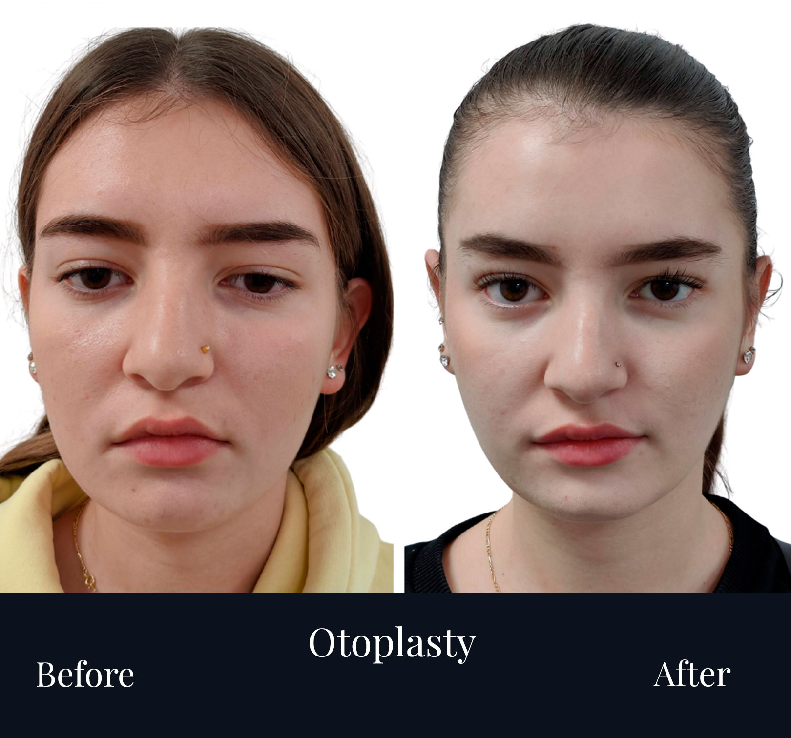
Ear Surgery (Otoplasty)
Prominent ears can draw attention away from the face, especially on front and three-quarter views. Otoplasty rebalances the ear by restoring natural folds and reducing projection—subtly, symmetrically, and with scars concealed behind the ear. I use cartilage-sparing, suture-based techniques to keep the helix and antihelix crisp while preserving a soft, natural rim.
What otoplasty can address
- Excess projection of the ear from the skull (open auriculo-mastoid angle)
- Under-defined antihelical fold giving a smooth, rounded ear
- Deep or over-projected conchal bowl pushing the ear outward
- Lobule prominence or asymmetry between ears
- Residual deformity after trauma or previous surgery
Suitable for adolescents and adults once ear growth is essentially complete; candidacy is confirmed in consultation.
My planning framework
Your evaluation includes standardised photography and measurements of auriculo-mastoid angle, helical-rim position, conchal depth, and lobule vector. We also review skin type (keloid risk), hairline, glasses/headwear habits, and sleeping position. The plan is individualised to avoid over-correction and the “pinned-back” look.
Technique
(tailored to your anatomy)
- Anaesthesia & setting: local anaesthesia with light sedation or general, in an operating theatre
- Incision: hidden in the posterior auricular crease (behind the ear)
- Cartilage shaping:
- Antihelix definition using permanent, concealed sutures to recreate the natural fold
- Conchal setback with sutures to reduce outward projection when the bowl is deep
- Lobule refinement when the lower pole contributes to prominence
- Closure: layered sutures; a soft protective dressing is placed at the end
These manoeuvres keep the ear’s natural curves while bringing it closer to the head in a controlled, symmetric way.
Recovery & aftercare
- Day 0–2: head elevation; mild pressure sensation is common
- Dressing: initial bulky dressing removed at the first review; then a soft headband day & night for 7–10 days, nights only for ~1 month
- Work/school: usually 5–7 days off (individual variation)
- Exercise: avoid impact, swimming, and steam rooms for ~4 weeks; no contact sports until cleared
- Sleeping: on your back to protect sutures during the early phase
Most swelling and bruising settle in 10–14 days; fine refinement continues as tissues relax over several weeks.
Results & longevity
Expect ears that sit closer to the head with a defined antihelical fold and a natural helical rim—not flat or over-tightened. Results are long-lasting; sutures are permanent to maintain fold definition.
Risks & how I minimise them
Common, usually mild: swelling, bruising, temporary numbness, suture irritation.
Less common: haematoma (requires urgent attention), infection, hypertrophic or keloid scarring (we stratify risk and use early scar care), asymmetry or partial relapse, contour irregularities. Conservative, layered techniques and structured follow-up reduce these risks.
Who is a good candidate?
- Stable health, non-smoker or willing to pause smoking around surgery
- Realistic goals: refinement and symmetry, not a “stuck-on” appearance
- Willing to follow headband and sleep-position guidance during healing
FAQs
Will there be visible scars?
Scars sit in the fold behind the ear and are typically discreet once mature.
Can otoplasty change my hearing?
No—the surgery reshapes external cartilage; hearing is unaffected.
Is cartilage removed?
In many cases we prefer suture-based reshaping; limited cartilage contouring is used only when necessary for projection control.
What about keloid risk?
We discuss your skin type and history. Early scar care (silicone, sun protection) and close follow-up help mitigate risk; steroid therapy is available if indicated.
How soon can I wear glasses or earphones?
Glasses may rest gently after the first review; avoid pressure on the suture line until advised. In-ear headphones are usually fine; over-ear models may need to wait.
Results